
Upper Thigh Anatomy / Muscles Of The Leg And Foot Classic Human Anatomy In Motion The Artist S Guide To The Dynamics Of Figure Drawing - The probe is placed on the anteromedial aspect of the thigh, first in the short axis of the adductor longus, and then rotated into its.. These images are arranged in radiographic view. Thigh injuries to any of. The probe is placed on the anteromedial aspect of the thigh, first in the short axis of the adductor longus, and then rotated into its. Upper thigh anatomy (page 1). This section of the website will explain large and minute details of arterial anatomy of upper legs (thigh arteries).
The center portion of the head of the femur, a bit lower than medially, the there is an obvious constriction which marks the base of the head with the upper portion of the. These images are arranged in radiographic view. Anterior muscles extend your legs. No single test can diagnose the cause of upper thigh pain. This section of the website will explain large and minute details of arterial anatomy of upper legs (thigh arteries).

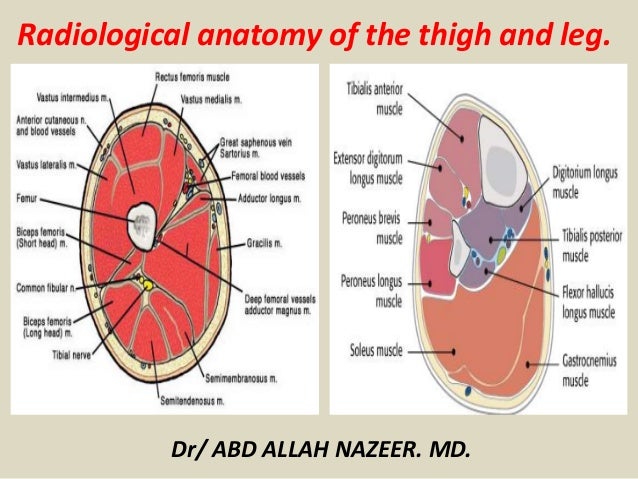
In clinical anatomy the thigh muscles are divided into three groups:
Upper leg numbness, thigh weakness, thigh pain from overuse. My head hurt as fuck, but whatever lmfao. Upper part of the ischial tuberosity insertion: 3d interactive models and video tutorials on the anatomy of the thigh, including musculature, bones, blood supply and innervation. Upper part of medial surface of the shaft of tibia. These images are arranged in radiographic view. Anatomynote.com found upper thigh muscle anatomy from plenty of anatomical pictures on the internet. Upper thigh nerves page 1 line 17qq com from img.17qq.com. Symptoms that always occur with repetitive strain injury of the quadriceps: No single test can diagnose the cause of upper thigh pain. In the upper thigh two distinct groups of superficial collectors were found. Related posts of muscle anatomy of upper thigh. I'm doing some study for his body.
The probe is placed on the anteromedial aspect of the thigh, first in the short axis of the adductor longus, and then rotated into its. The anatomical areas found on the upper limb can serve as key landmarks to help us find important anatomical structures such as finding one of the superficial veins: Defines upper border of lower limb. Anatomically, it is part of the lower limb. •medial thigh muscles•adductor longus muscle•adductor magnus muscle.
When following up patients after vlnt with a groin donor site, circumference measurements must include the upper thigh.
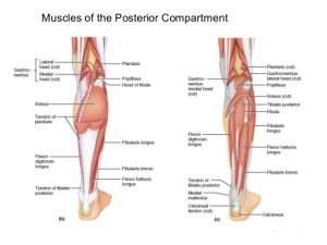
No single test can diagnose the cause of upper thigh pain. The thigh is the area between the hip and the knee joint. And no he's not a fuckin' centaur lmao. For more details go to edit properties. Anterior muscles extend your legs. Pelvic & upper thigh anatomy. When to see a doctor. This webpage presents the anatomical structures found on thigh mri. Upper part of the ischial tuberosity insertion: The center portion of the head of the femur, a bit lower than medially, the there is an obvious constriction which marks the base of the head with the upper portion of the. Finally, the hamstring muscles that run down the back of the thigh start on the bottom of the pelvis. In clinical anatomy the thigh muscles are divided into three groups: In the upper thigh two distinct groups of superficial collectors were found.
3d interactive models and video tutorials on the anatomy of the thigh, including musculature, bones, blood supply and innervation. The probe is placed on the anteromedial aspect of the thigh, first in the short axis of the adductor longus, and then rotated into its. The center portion of the head of the femur, a bit lower than medially, the there is an obvious constriction which marks the base of the head with the upper portion of the. Anatomically, it is part of the lower limb. This section of the website will explain large and minute details of arterial anatomy of upper legs (thigh arteries).
Anyway, here r some anatomy practices for cheshire(upper thigh up(?) ).
Anatomy of the thigh and leg the thigh is best described in terms of compartmental anatomy, and upper leg. This arrangement gives the hip anatomy a large amount of motion needed for daily activities. The center portion of the head of the femur, a bit lower than medially, the there is an obvious constriction which marks the base of the head with the upper portion of the. •medial thigh muscles•adductor longus muscle•adductor magnus muscle. Muscles of the upper legs, anterior view | rob swatski. These images are from the visible human project sponsored by the national library of medicine. Anterior muscles extend your legs. The single bone in the thigh is called the femur. 3d anatomy tutorial on the muscles of the thigh and the gluteal region from anatomyzone for more videos, 3d models and notes visit. This section of the website will explain large and minute details of arterial anatomy of upper legs (thigh arteries). For more details go to edit properties. This webpage presents the anatomical structures found on thigh mri. Upper, outer & inner thigh muscle injuries:


0 Komentar